
Discover the causes, symptoms, and treatment of Epidermal Necrosis. Learn how to manage this skin condition effectively in our comprehensive guide.
Did you know that toxic epidermal necrolysis, a severe form of epidermal necrolysis, has a mortality rate of approximately 25-30% according to Johns Hopkins Medicine? This life-threatening condition, often triggered by drug reactions, affects thousands worldwide each year.
Epidermal necrosis, including its more severe form, toxic epidermal necrolysis, is a critical skin disorder that requires immediate medical attention. It is primarily caused by adverse drug reactions, with medications like antibiotics and anticonvulsants being common culprits. Understanding this condition is vital for both patients and healthcare providers to prevent complications and improve outcomes.
Epidermal necrolysis is characterized by widespread skin damage, leading to detachment of the epidermis from the dermis. This can result in severe complications, including infections and organ failure, if not managed promptly. Proper drug management and early diagnosis are crucial in avoiding these adverse outcomes.
Differentiating epidermal necrolysis from other skin conditions is essential for accurate treatment. This guide will explore the causes, symptoms, diagnosis, treatment, and prevention measures associated with epidermal necrolysis, providing a comprehensive understanding for those affected and their caregivers.
Key Takeaways
- Toxic epidermal necrolysis is a life-threatening condition with significant mortality rates.
- Drug reactions are the primary cause of epidermal necrolysis, emphasizing the need for careful medication monitoring.
- Early diagnosis is critical to prevent severe complications and improve patient outcomes.
- Understanding the differences between epidermal necrolysis and other skin conditions is vital for effective treatment.
- This guide provides comprehensive insights into causes, symptoms, diagnosis, treatment, and prevention of epidermal necrolysis.
Understanding Epidermal Necrosis
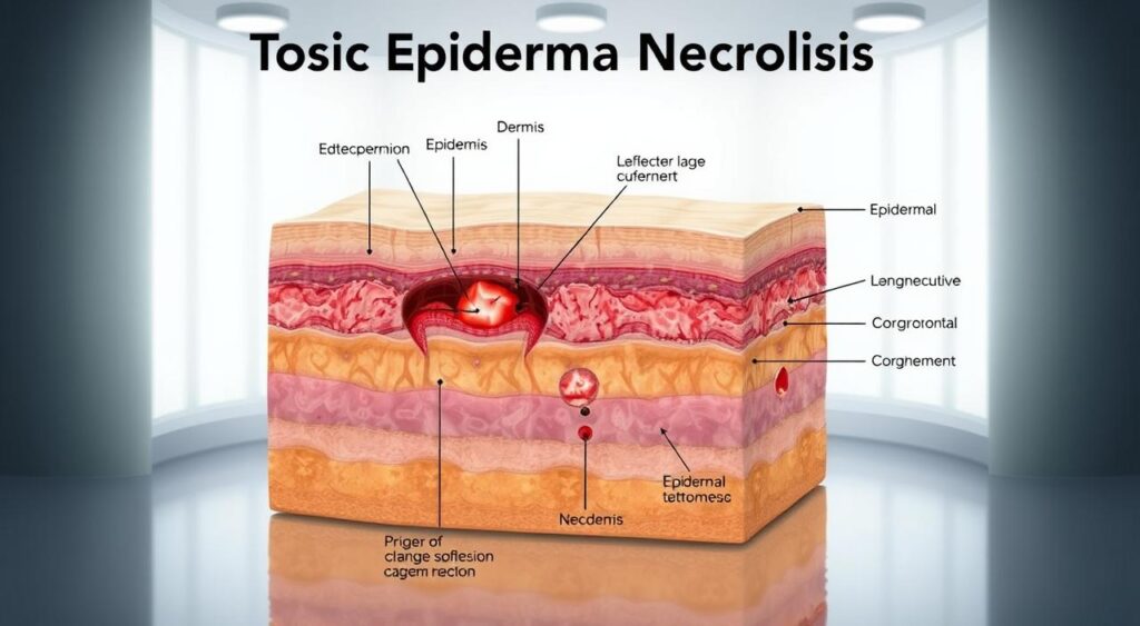
Epidermal necrosis is a severe skin condition characterized by the detachment of the epidermis from the dermis, leading to full-thickness skin damage. It often involves mucosal surfaces and can be life-threatening if not treated promptly.
Defining the Condition
The condition arises from pathological processes that cause extensive skin cell death. This damage can result from drug reactions, infections, or other systemic factors. The detachment of the epidermis from the dermis creates a vulnerable environment, increasing the risk of infections and other complications.
How It Differs from Related Syndromes
Epidermal necrosis is often compared to Stevens-Johnson syndrome (SJS), but the two differ in severity and triggers. SJS typically involves less skin detachment and is more commonly linked to drug reactions. In contrast, epidermal necrosis can be caused by a broader range of factors, including severe infections, and results in more extensive skin damage.
While both conditions share similar symptoms, such as skin lesions and mucosal involvement, epidermal necrosis is generally more severe. Early diagnosis is crucial to prevent complications and improve patient outcomes.
Causes and Risk Factors Behind Toxic Epidermal Necrolysis
Toxic epidermal necrolysis (TEN) is primarily triggered by drug reactions, infections, and genetic predispositions. Understanding these factors is crucial for prevention and early intervention.
Drug-Induced Reactions and Other Triggers
Adverse drug reactions are the leading cause of TEN, particularly with medications like anticonvulsants and antibiotics. These drugs can provoke severe immune responses.
Besides drugs, infections and environmental factors can also trigger TEN. For instance, viral infections may sometimes initiate the condition.
Genetic and Immune System Factors
Genetic predispositions, such as specific HLA alleles, increase the risk of developing TEN. These genetic markers can affect how the immune system responds to certain drugs.
The immune system plays a significant role in TEN. T-cell activation leads to keratinocyte apoptosis, causing skin damage and detachment.
| Cause | Description | Risk Level |
|---|---|---|
| Drug Reactions | Medications like anticonvulsants and antibiotics | High |
| Genetic Factors | Specific HLA alleles | Medium |
| Immune Response | T-cell activation leading to apoptosis | High |
These factors often work together, leading to widespread skin involvement and severe complications if not addressed promptly.
Identifying Symptoms and Early Signs
Recognizing the early signs of toxic epidermal necrolysis is crucial for timely intervention. The condition often begins with nonspecific symptoms that can progress rapidly, making early diagnosis challenging but vital.
Prodromal Flu-like Symptoms
The initial stage of TEN often mimics common illnesses, with symptoms such as fever, fatigue, and sore throat. These prodromal symptoms can last for 1-3 days before skin changes appear. In some cases, patients may experience coughing or conjunctivitis, further complicating early diagnosis.
Skin and Mucosal Changes
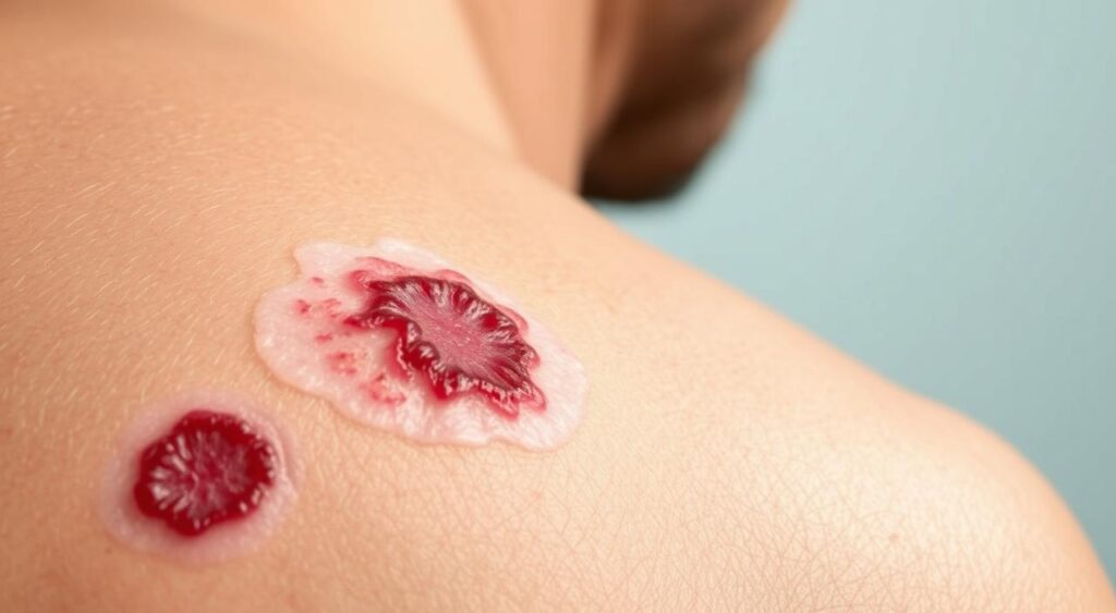
The skin manifestations typically begin with painful erythematous macules and papules, which can rapidly progress to blisters and peeling. Mucous membranes are particularly affected, with the eyes, mouth, and genital areas being highly vulnerable. These changes can lead to severe discomfort and functional impairment.
| Stage | Description | Timeline |
|---|---|---|
| Prodrome | Flu-like symptoms such as fever and fatigue | 1-3 days |
| Early Skin Changes | Appearance of painful blisters and erythematous lesions | 2-4 days after prodrome |
| Severe Skin Detachment | Widespread skin peeling and mucosal involvement | 4-7 days from onset |
Early recognition of these signs is critical to prevent life-threatening complications. If you or someone you know experiences these symptoms, seek immediate medical attention to ensure prompt diagnosis and treatment.
Diagnosing Epidermal Necrosis
Accurate diagnosis of epidermal necrosis requires a combination of clinical evaluation and diagnostic tests. Early detection is critical to prevent severe complications and improve patient outcomes.
Clinical Evaluations and Physical Examinations
During the clinical evaluation, healthcare providers assess the extent of skin detachment and check for signs like the Nikolsky sign, where gentle pressure on the skin causes it to separate. They also examine mucous membranes, particularly in the eyes, mouth, and genital areas, for signs of involvement.
Biopsy and Histologic Confirmation
A skin biopsy is essential for confirming the diagnosis. Histologic examination reveals full-thickness epidermal necrosis, distinguishing it from other conditions like Stevens-Johnson syndrome (SJS). The biopsy results help guide treatment and prognosis.
| Diagnostic Method | Description | Importance |
|---|---|---|
| Clinical Evaluation | Assessment of skin detachment and mucous membrane involvement | Identifies severity and affected areas |
| Skin Biopsy | Confirms full-thickness epidermal necrosis | Distinguishes from other conditions like SJS |
| SCORTEN Score | Predicts mortality risk based on clinical factors | Guides treatment and prognosis |
Using tools like the SCORTEN scoring system helps predict mortality risk, ensuring timely and appropriate interventions. Rapid diagnosis is vital to enhance patient outcomes in this severe disorder.
Treatment Options and Patient Management
Effective management of severe skin conditions like toxic epidermal necrolysis requires a multi-faceted approach. Immediate hospitalization is critical, especially in specialized units such as intensive care or burn centers, where patients can receive the necessary supportive care and monitoring.
Hospitalization and Supportive Care
Hospitalization ensures that patients receive round-the-clock care. Supportive measures include IV fluids to prevent dehydration, wound care to promote healing, and pain management to keep patients comfortable. These steps are essential for stabilizing the patient and preventing further complications.
Medications and Alternative Therapies
Systemic medications like IV immunoglobulins and corticosteroids are often used, though their effectiveness can vary. Alternative therapies, such as those targeting the immune system, are being explored to modulate the body’s response and reduce skin damage. Early withdrawal of any offending medications is crucial to halt the progression of the condition.
| Treatment Type | Details | Effectiveness |
|---|---|---|
| Supportive Care | Includes IV fluids and wound management | High |
| Systemic Medications | IV immunoglobulins and corticosteroids | Moderate |
| Alternative Therapies | Immune system modulation | Emerging |
Early intervention and comprehensive patient management are vital to improving outcomes and reducing the risk of long-term complications.
Prognosis and Potential Complications
The prognosis for individuals with toxic epidermal necrolysis (TEN) varies widely, depending on several factors, including the severity of skin detachment and the patient’s overall health. Understanding prognostic scores and long-term effects is crucial for managing the condition effectively.
Understanding Prognostic Scores
The SCORTEN score is a widely used prognostic tool for TEN. It assesses factors such as age, heart rate, and the extent of skin detachment to predict mortality rates. A higher SCORTEN score indicates a poorer prognosis. For instance, a score of 3 or more is associated with a mortality rate of approximately 90%.
| SCORTEN Component | Details | Impact on Prognosis |
|---|---|---|
| Age | Patients over 40 years | Higher mortality risk |
| Heart Rate | Increased heart rate | Indicates severe stress response |
| Skin Detachment | Extent of skin involvement | Greater detachment worsens prognosis |
Long-Term Effects on Skin and Mucosal Health
Survivors often face chronic issues, such as skin scarring and ocular problems. The spectrum of long-term effects includes persistent blistering and pigment changes, which can significantly impact quality of life. In severe cases, the peel process may lead to permanent disfigurement.
| Long-Term Effect | Description | Prevalence |
|---|---|---|
| Chronic Scarring | Persistent skin damage | Common in severe cases |
| Ocular Issues | Vision problems | High in advanced TEN |
| Pigment Changes | Discoloration | Variable |
Early intervention is critical to improve outcomes and reduce complications. The spectrum of TEN’s severity underscores the need for vigilant monitoring and prompt treatment.
Preventing Severe Outcomes in Epidermal Necrosis
Preventing severe outcomes in cases of epidermal necrosis requires a proactive approach. Early identification of causative medications is crucial to halt the progression of the condition. Prompt withdrawal of the offending drug can significantly improve patient outcomes.
Early Medication Withdrawal
One of the most critical steps in managing epidermal necrosis is the immediate cessation of any medication suspected to be the cause. Studies have shown that early withdrawal can prevent further skin detachment and reduce the risk of life-threatening complications. Healthcare providers should maintain a high index of suspicion for drug-related reactions, especially in patients on new medications.
Strategies for Patient and Clinical Care
Clinical monitoring should be intensified in patients at high risk. This includes regular skin assessments and monitoring for signs of mucosal involvement. Patient education plays a vital role in early detection. Educating patients on recognizing early warning signs, such as unusual rashes or mucosal lesions, can lead to timely medical intervention.
Genetic screening may also be recommended for individuals with a family history of severe cutaneous adverse reactions. This can help identify those at higher risk, allowing for closer monitoring and personalized management plans.
Research emphasizes the importance of rapid intervention and proactive management. Multidisciplinary care, involving dermatologists, immunologists, and other specialists, ensures comprehensive patient management. Coordinated care can significantly reduce severe outcomes and improve long-term recovery.
Conclusion
In conclusion, managing severe cutaneous adverse reactions like Stevens-Johnson syndrome and toxic epidermal necrolysis demands a comprehensive approach. These conditions, often triggered by drug reactions, can lead to life-threatening complications if not addressed promptly. Early diagnosis and aggressive treatment are vital to improving patient outcomes and reducing mortality rates.
Understanding the causes, symptoms, and treatment options is essential for both patients and healthcare providers. This guide has outlined the complexities of these conditions, emphasizing the need for clinical vigilance and ongoing research. By staying informed and proactive, we can enhance patient care and reduce the impact of severe cutaneous adverse reactions.
If you suspect any symptoms of these conditions, seek immediate medical attention. Consulting trusted healthcare professionals is crucial for personalized advice and effective management. Together, we can raise awareness and improve outcomes for those affected by these severe skin disorders.
FAQ
Q: What is toxic epidermal necrolysis (TEN)?
A: TEN is a severe skin disorder triggered by certain medications. It leads to widespread skin death and detachment, often resembling burns. Prompt medical attention is critical to manage this life-threatening condition.
Q: How does TEN differ from Stevens-Johnson Syndrome (SJS)?
A: TEN and SJS are part of the same spectrum of severe cutaneous adverse reactions. SJS involves less skin detachment (typically under 10%) and more mucosal involvement, while TEN involves more widespread skin loss (over 30%) and is often more severe.
Q: What are the most common symptoms of TEN?
A: Symptoms include painful skin blisters, peeling, and redness, often starting in the mucous membranes (eyes, mouth, genitals). Flu-like symptoms may precede skin changes, and patients may experience eye inflammation or vision issues.
Q: What medications commonly cause TEN?
A: Anticonvulsants, antibiotics (like sulfonamides), and nonsteroidal anti-inflammatory drugs (NSAIDs) are frequent culprits. The reaction is rare but can occur within days to weeks of starting a new medication.
Q: How is TEN diagnosed?
A: Diagnosis involves a clinical examination, patient history review, and sometimes a skin biopsy. Histologic confirmation helps distinguish TEN from other conditions like burns or infections.
Q: What is the standard treatment for TEN?
A: Treatment focuses on supportive care, often in a burn unit or ICU. This includes wound management, fluid replacement, and stopping the offending medication. In some cases, medications like intravenous immunoglobulin (IVIG) may be used.
Q: Can TEN be fatal?
A: Yes, TEN can be life-threatening, with mortality rates ranging from 20% to 50%. Timely intervention and proper care are crucial to improve survival rates and reduce long-term complications.
Q: How can TEN be prevented?
A: Early recognition of symptoms and immediate withdrawal of the suspected medication are key. High-risk patients should be closely monitored when starting new drugs, especially those known to cause severe reactions.
Q: What are the long-term effects of TEN?
A: Survivors may experience chronic skin issues, vision problems, and psychological trauma. Regular follow-ups with dermatologists and ophthalmologists are essential for managing these complications.
Q: Are there support groups for TEN patients?
A: Yes, organizations like the Stevens-Johnson Syndrome Foundation and online forums provide resources and support for patients and families affected by TEN and related conditions.