
This advanced imaging method is key in diagnosing and treating heart and artery issues. It lets doctors see blood vessels live, helping them make better treatment plans.
In this guide, we’ll dive into angiography’s basics, its history, and its uses in medicine today. We’ll cover how contrast agents work and the latest tech in this field. You’ll get a full picture of this cutting-edge imaging method.
Key Takeaways
- Angiography is a vital imaging technique that uses X-rays and dye to see blood vessels.
- It’s crucial for diagnosing and treating heart and artery problems.
- Angiography has grown, using new catheter techniques and digital subtraction.
- It’s used for both checking and treating conditions, like in interventional radiology.
- Keeping patients safe and protecting them from radiation is key in angiography.
Understanding Medical Imaging and Its Evolution
Medical imaging has changed a lot in the last 100 years. It started with X-rays and now we have diagnostic imaging that’s much better. This change has helped doctors care for patients better, make more accurate diagnoses, and create treatment plans that fit each person.
Historical Development of Diagnostic Imaging
In 1895, Wilhelm Röntgen from Germany found X-rays. This was a big step in medicine, letting doctors see inside the body for the first time.
After that, X-ray imaging grew with new tech like fluoroscopy, CT scans, and MRI. These tools helped doctors understand the body better and find diseases more easily.
From X-rays to Modern Imaging Solutions
Now, diagnostic imaging includes many types, like ultrasound and PET scans. These methods give detailed views of the body and help doctors understand how it works. This helps them make better choices for patient care.
Digital technology has made medical imaging even better. It makes images faster, clearer, and easier to manage. AI is also changing things, making diagnosis faster and improving care.
The future of diagnostic imaging looks bright. We can expect more advances in things like molecular imaging and personalized medicine. These changes will help doctors help patients even more.
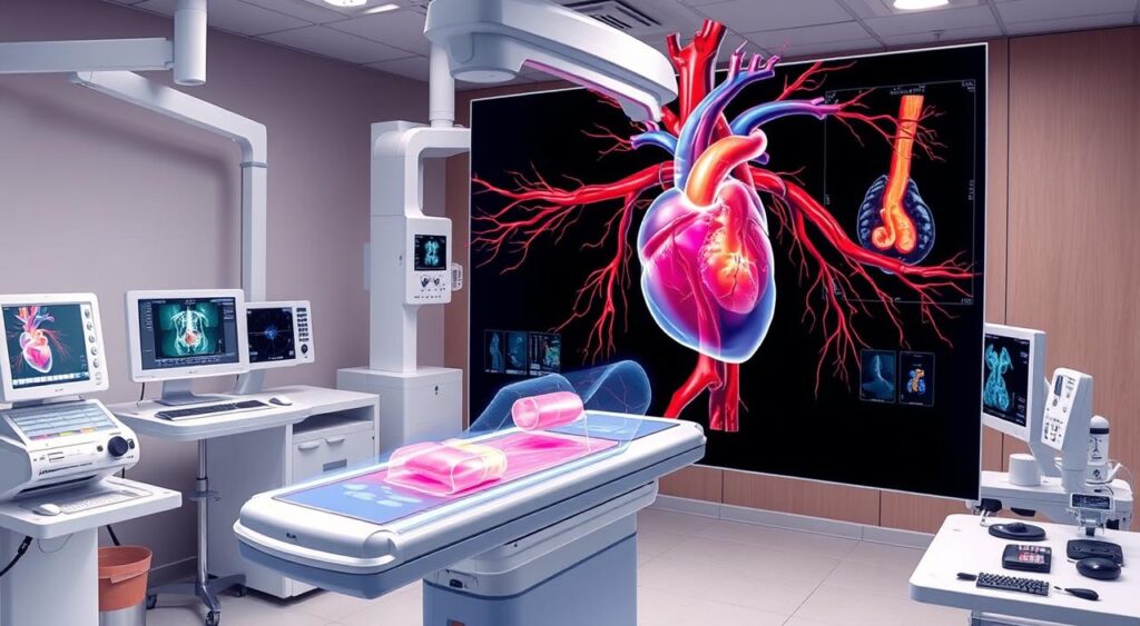
What is Angiography: Core Principles and Functions
Angiography is a cutting-edge medical imaging method. It helps doctors see the blood vessels in our bodies. This non-invasive test is key for diagnosing and treating heart and blood vessel problems.
At its heart, angiography uses special tools to take detailed pictures of blood vessels, or the vascular system. A contrast dye is injected into the blood. This makes it easy to see the arteries, veins, and capillaries that move blood around our body.
The main jobs of angiography include:
- Finding and checking for blockages, narrow spots, or other issues in blood vessels
- Spotting the exact spot and size of diseases like atherosclerosis or aneurysms
- Helping with procedures like angioplasty or stent placement to improve blood flow
- Looking at how well the heart and blood vessels are working
Angiography gives a clear view of our blood vessel visualization. It’s a key tool in angiography and heart care today.
| Angiographic Technique | Description |
|---|---|
| Conventional Angiography | Uses a catheter to inject a contrast dye directly into the blood vessels, allowing for detailed x-ray imaging of the vascular system. |
| Computed Tomography Angiography (CTA) | Utilizes a specialized CT scan to capture high-resolution, three-dimensional images of the blood vessels, often used for evaluating the coronary arteries. |
| Magnetic Resonance Angiography (MRA) | Employs magnetic resonance imaging (MRI) technology to generate images of the blood vessels without the use of ionizing radiation. |
Knowing how angiography works helps doctors give better care. They can manage many heart and blood vessel problems more effectively.
“Angiography is a powerful diagnostic tool that allows us to see the intricate details of the body’s vascular system, enabling us to detect and address a variety of cardiovascular and circulatory issues with greater precision and effectiveness.”
Types of Angiographic Procedures in Modern Medicine
Angiography is a key tool in modern medicine. It helps doctors see blood vessels clearly. This lets them check the heart and brain’s blood flow. There are three main types: coronary, cerebral, and peripheral angiography.
Coronary Angiography
Coronary angiography looks at the heart’s blood vessels. It helps find blockages or narrowings that can lead to heart disease. Doctors use a dye and special cameras to see the heart’s blood vessels.
Cerebral Angiography
Cerebral angiography looks at the brain’s blood vessels. It’s great for finding problems like strokes or aneurysms. Doctors use dye and cameras to see the brain’s blood flow.
Peripheral Angiography
Peripheral angiography looks at blood vessels outside the heart and brain. It’s used to find problems in the limbs. Doctors use dye and cameras to see the blood flow in arms and legs.
These angiographic procedures are vital in modern medicine. They help doctors diagnose and treat many heart and brain problems. This improves patient care and outcomes.
| Angiographic Procedure | Target Area | Key Applications |
|---|---|---|
| Coronary Angiography | Heart’s blood vessels (coronary arteries) | Diagnosis and management of heart disease, including blockages and narrowing of the arteries |
| Cerebral Angiography | Blood vessels in the brain | Evaluation and treatment of cerebrovascular conditions, such as strokes, aneurysms, and arteriovenous malformations |
| Peripheral Angiography | Blood vessels in the limbs (arms and legs) | Diagnosis and management of peripheral artery disease (PAD) and other vascular abnormalities in the extremities |
These angiographic procedures are key in modern medicine. They help doctors diagnose and treat many heart and brain problems. This improves patient care and outcomes.
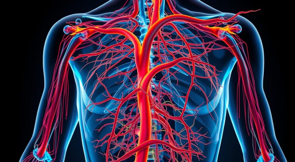
The Role of Contrast Agents in Blood Vessel Visualization
Angiography is a key tool in modern medicine. It uses contrast agents to see blood vessels clearly. These substances help doctors get detailed images for accurate diagnosis and treatment.
Contrast agents are injected into the blood. They make blood vessels more visible on scans like X-rays and MRIs. This helps doctors spot problems like blockages or abnormalities.
There are different types of contrast agents. Iodine-based agents are often used in coronary angiography. Gadolinium-based agents are better for MRI scans because they show soft tissues well.
Using contrast agents is a careful process. Doctors consider the patient’s health and any allergies. They choose the right agent and dose to avoid bad reactions and get clear images.

Contrast agents help doctors understand blood vessel health. This knowledge helps manage many heart conditions. It leads to better patient care and quality of life.
Advanced Catheterization Techniques in Angiography
Angiographic procedures use special catheters to get detailed images of the body’s blood vessels. This technique is key in catheterization and interventional radiology.
Catheter Selection and Navigation
Choosing the right catheter is crucial for angiographic exams. Radiologists pick catheters based on the procedure’s needs and the patient’s body. They use their skills to guide these catheters through the blood vessels safely and effectively.
Patient Preparation and Safety Protocols
Keeping patients safe is a big deal in catheterization and interventional radiology. Before the exam, patients get checked and told what to do. The team follows strict safety rules, like using clean techniques and watching the patient’s health closely.
| Catheter Type | Commonly Used for | Key Features |
|---|---|---|
| Pigtail Catheter | Aortic and cardiac imaging | Curved shape for easy access to the heart and aorta |
| Diagnostic Catheter | General vascular imaging | Straight or slightly curved design for versatile use |
| Guiding Catheter | Interventional procedures | Larger diameter for accommodating therapeutic devices |
“Precise catheter selection and navigation, combined with meticulous patient preparation, are essential for successful angiographic examinations and subsequent interventional procedures.”
Digital Subtraction Angiography (DSA) Technology
In the world of cardiovascular imaging, angiography has been key. But Digital Subtraction Angiography (DSA) has changed the game. It offers better views and a simpler way to check blood vessels. This new tech is a big deal in angiography and cardiovascular imaging.
DSA uses digital tech to show blood vessels clearly. It removes background images, giving a sharp view of the blood vessels. This helps doctors see the heart’s system better, making their decisions more accurate.
DSA is great at showing small blood vessels clearly. This is very important for finding and treating heart problems. It’s a key tool in modern medicine for looking at the heart and blood vessels.
| Feature | Benefit |
|---|---|
| Enhanced Contrast Visualization | Improved detection and diagnosis of vascular abnormalities |
| Reduced Radiation Exposure | Increased patient safety during diagnostic procedures |
| Real-Time Imaging Capabilities | Enables precise guidance during interventional procedures |
| Advanced Image Processing Algorithms | Enhances the clarity and quality of angiographic images |
As angiography and cardiovascular imaging grow, DSA’s role is more important. It gives doctors a clear view and precision. This has changed how they treat heart and blood vessel problems, leading to better care for patients.
Interventional Radiology Applications in Angiographic Procedures
Angiography is key in interventional radiology. It helps treat many heart problems without big surgery. This method uses special images to guide treatments, making them more precise and effective.
Therapeutic Interventions
Angiography is used to treat heart diseases. It helps place stents and open blocked blood vessels. This way, doctors can fix problems without making a big cut, which means patients heal faster and face fewer risks.
Emergency Applications
Angiography is also vital in emergency care. It helps quickly find and fix serious issues like aortic dissections and pulmonary embolisms. This fast action can save lives by stopping severe problems before they get worse.
The mix of angiography and interventional radiology has changed how we treat heart problems. It gives patients better, more focused care that’s less invasive.
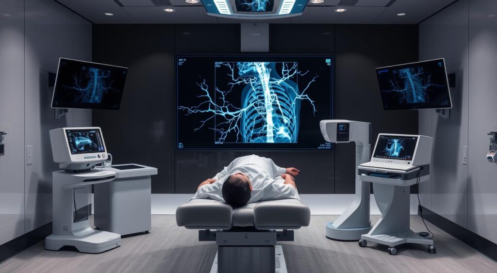
Fluoroscopy and Real-time Imaging During Procedures
Fluoroscopy is a key tool in modern angiographic procedures. It lets doctors see blood vessels in real-time. This helps them use catheters and guide treatments safely and accurately. It’s especially useful in fluoroscopy and cardiovascular imaging.
During angiographic procedures, fluoroscopy helps doctors watch catheters and contrast agents move. This feedback lets them make quick, precise adjustments. It reduces the chance of problems and boosts the success of treatments.
Fluoroscopy uses X-rays to create images. These images are shown on a screen in real-time. This lets doctors see the body’s internal structures, like blood vessels, as they move.
| Fluoroscopy Advantages | Fluoroscopy Limitations |
|---|---|
| Real-time, continuous visualization of blood vessels Improved navigation and guidance of catheters and instruments Enhanced accuracy and safety of interventional procedures | Exposure to ionizing radiation Potential for tissue damage with prolonged exposure Limited depth of penetration compared to other imaging modalities |
Healthcare providers follow strict safety rules to use fluoroscopy safely. They use protective gear to lower radiation exposure. This careful approach ensures fluoroscopy is used wisely and effectively.
“Fluoroscopy has revolutionized the way we approach cardiovascular interventions, allowing us to see the inner workings of the body in real-time and guide our treatments with unprecedented precision.”
Patient Safety and Radiation Protection Measures
In angiography and interventional radiology, patient safety is key. These procedures use ionizing radiation, so reducing exposure is vital for healthcare workers.
Minimizing Radiation Exposure
Healthcare professionals take many steps to lower radiation risks. They use:
- Optimizing X-ray settings to use less radiation.
- Collimation to focus the beam on the needed area.
- Fluoroscopic imaging for real-time views with less radiation.
- Dose-reduction methods like pulsed fluoroscopy and shorter exposure times.
Protective Equipment and Protocols
Healthcare teams and patients also follow strict safety rules. They use special gear:
| Protective Equipment | Purpose |
|---|---|
| Lead aprons and thyroid shields | Shielding vital organs from radiation |
| Leaded eyeglasses or face shields | Protecting the eyes and face |
| Radiation monitoring devices | Tracking and recording radiation exposure |
These steps, along with staff training and quality checks, keep patients safe during angiography and interventional radiology procedures.
“Radiation safety is a critical aspect of patient care in the field of angiography and interventional radiology. Adhering to strict protocols and utilizing protective equipment helps minimize the risks associated with these essential diagnostic and therapeutic procedures.”
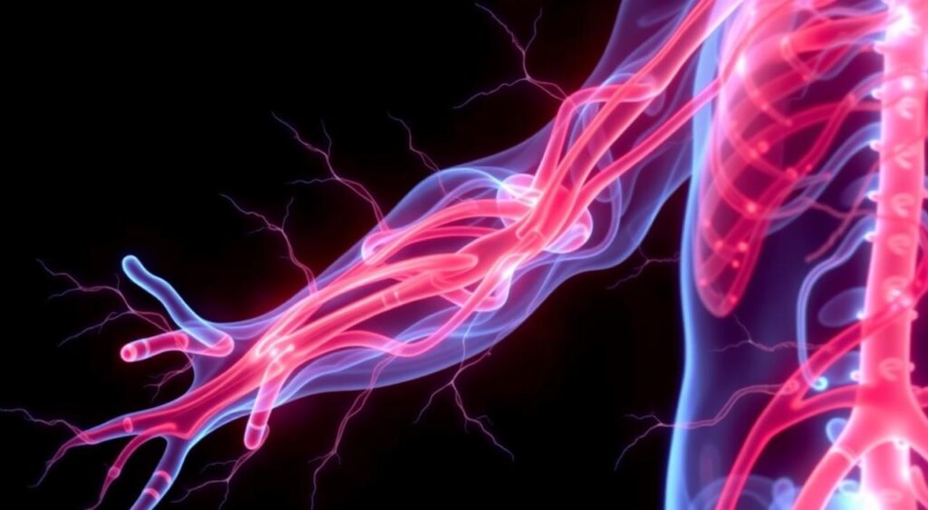
Diagnostic Applications in Cardiovascular Disease
Angiography is a key tool in diagnosing heart and blood vessel problems. It lets doctors see the blood vessels clearly. This helps them spot blockages and other issues.
It’s mainly used to find cardiovascular disease. Doctors use a contrast agent to see the blood vessels and heart. This helps them plan the best treatment.
Coronary arteriography is crucial for coronary artery disease. It shows blockages in the heart’s arteries. This helps doctors fix the problem before it gets worse.
Cerebral angiography helps with brain problems like stroke. It shows the brain’s blood vessels. This helps doctors find and fix the issue.
Angiography also helps with peripheral artery disease. It checks the arteries in the legs and arms. This helps find blockages that can cause pain and weakness.
In short, angiography is very important in heart and blood vessel care. It helps doctors make the right diagnosis and treatment plans. This improves patient care for many heart and blood vessel problems.
“Angiography is a vital diagnostic tool that allows us to see the intricate network of blood vessels with remarkable clarity, enabling us to identify and address a wide range of cardiovascular issues.”
Preparing for an Angiographic Examination
Going through an angiographic procedure is a big step in your medical journey. It’s important to prepare well for a good outcome. Knowing what to do before and after can make things easier and less stressful.
Pre-procedure Guidelines
Your doctor will give you instructions before the angiography. These might include:
- Fasting for 6-8 hours to have an empty stomach
- Stopping certain medications, like blood thinners, as told
- Getting someone to drive you home because of possible side effects
- Telling your doctor about any allergies, especially to iodine or contrast dyes
Recovery and Aftercare
After the procedure, you’ll be watched in a recovery area. The catheter will be taken out, and a bandage or compression device will be put on the site. It’s key to follow your doctor’s advice on rest, drinking fluids, and any medication.
You can usually go back to normal activities in a day or two. But, you should avoid hard exercise or heavy lifting for a few days. Your doctor will tell you when it’s okay to go back to your usual life.
Being well-prepared and taking good care of yourself can make your angiographic exam and recovery smoother. By knowing what to do before and after, you can feel more in control and confident during your healthcare journey.
Latest Technological Advancements in Angiographic Imaging
In the world of heart imaging, angiography has seen big changes. These updates have made it easier and safer to see blood vessels. They also make procedures more efficient.
Improving Imaging Quality
New high-resolution digital detectors are a big step forward. They work with advanced software to show blood vessels clearly. This helps doctors make better decisions for their patients.
Reducing Radiation Exposure
Keeping radiation low during tests is very important. New methods and software have made this possible. Now, patients and doctors get less radiation during these tests.
Enhancing Procedural Guidance
New imaging and navigation systems have changed how procedures are done. Techniques like 3D angiography and fusion imaging help doctors see better. This leads to better results and fewer problems.
| Technological Advancement | Benefit |
|---|---|
| High-resolution digital detectors | Improved image quality for more accurate diagnosis and treatment planning |
| Low-dose imaging techniques | Reduced radiation exposure for patients and medical staff |
| 3D rotational angiography and fusion imaging | Enhanced procedural guidance and improved outcomes |
As heart imaging keeps getting better, these new technologies will change how we treat blood vessel problems. This will lead to better care and outcomes for patients.
Common Complications and Risk Management
Angiography and interventional radiology are usually safe. But, there are risks that doctors need to be ready for. Knowing these risks and how to manage them is key to keeping patients safe and getting good results.
Bleeding is a big worry in angiography. It can happen at the site where the doctor inserts the catheter or inside the body. Choosing the right patient, using the right medicines, and being careful with the catheter can help lower this risk. Also, watching the patient closely and acting fast can help fix any bleeding problems.
- Allergic reactions to the contrast dye used in angiography are another risk. Checking for allergies, giving medicine before the test, and watching the patient closely can help avoid this.
- Vascular injuries, like dissection or perforation, can happen during the procedure. Using skilled interventional radiology techniques and watching the images closely is important to avoid and handle these problems.
- Embolization, where a clot or plaque blocks a blood vessel, is another risk. It’s important to spot and treat it quickly.
| Complication | Frequency | Risk Mitigation Strategies |
|---|---|---|
| Bleeding | 2-5% | Careful patient selection, medication management, and technique during catheter insertion |
| Allergic Reactions | 1-2% | Screening for allergies, premedication, and close patient monitoring |
| Vascular Injuries | 1-3% | Skilled interventional radiology techniques and continuous imaging guidance |
| Embolization | 1-2% | Prompt recognition and treatment |
To reduce these risks, doctors must follow strict safety rules, stay up-to-date with their skills, and be ready to act fast if problems come up. By focusing on patient safety and using good risk management, the benefits of angiography and interventional radiology can be fully enjoyed, while keeping the chance of bad outcomes low.
The Future of Angiographic Procedures
The field of cardiovascular imaging and interventional radiology is growing fast. New technologies and artificial intelligence (AI) in imaging analysis are changing how doctors diagnose and treat vascular conditions.
Emerging Technologies
New medical imaging tech is making angiographic procedures more precise and less invasive. Tools like dual-energy CT angiography and functional MRI show blood vessels and tissues better. This helps doctors find problems early and treat them more accurately.
Robotic-assisted angiography systems are also changing the game. They offer better control and precision, which can lower radiation exposure and improve patient results.
AI Integration in Imaging Analysis
AI in cardiovascular imaging is a big deal in interventional radiology. AI algorithms quickly analyze lots of data, spotting things humans might miss. This could make diagnoses more accurate and help doctors make faster decisions.
AI can also predict complications and help plan treatments. This means treatments can be more tailored to each patient. As these technologies get better, cardiovascular imaging will become more precise and focused on the patient.
“The future of angiographic procedures lies in the seamless integration of cutting-edge imaging technologies and the power of artificial intelligence. By harnessing these advancements, we can unlock new possibilities in the diagnosis and treatment of vascular diseases, ultimately enhancing patient outcomes and transforming the field of interventional radiology.”
Conclusion
Angiography has changed the game in modern medicine. It lets doctors diagnose and treat heart problems with great accuracy. This technique has come a long way from its early days in diagnostic imaging.
Now, angiography is a key tool in understanding our body’s blood vessels. It’s a non-invasive way to see how our blood flows. This helps doctors make better decisions for our health.
New technologies like digital subtraction angiography (DSA) have made angiography even better. These tools help doctors see more clearly and treat problems more effectively. This has changed how we deal with heart diseases.
Angiography is getting even better with the help of artificial intelligence. This means safer and more efficient treatments for patients. Doctors will soon be able to give care that’s more tailored to each person’s needs.
This will lead to better health outcomes for everyone. It’s an exciting time for cardiovascular healthcare. The future looks bright for patients and doctors alike.
FAQ
Q: What is angiography?
A: Angiography is a medical imaging method. It uses X-rays and contrast dye to see blood vessels. It’s key for diagnosing and treating heart and blood vessel problems.
Q: How does angiography work?
A: Angiography uses a contrast dye injected into the blood. This dye makes blood vessels visible under X-rays. Doctors can then check their structure and spot any issues.
Q: What are the different types of angiographic procedures?
A: There are several types of angiographic procedures. These include coronary, cerebral, and peripheral angiography. Each focuses on different parts of the body’s blood vessels.
Q: What is the role of contrast agents in angiography?
A: Contrast agents are vital in angiography. They make blood vessels stand out during imaging. These substances temporarily make blood denser, helping to see vessels clearly on X-rays.
Q: How are angiographic procedures performed?
A: Angiographic procedures start with a thin, flexible tube called a catheter. It’s inserted into a blood vessel, usually in the groin or arm. The catheter guides to the needed area, and dye is injected for real-time imaging.
Q: What is Digital Subtraction Angiography (DSA)?
A: Digital Subtraction Angiography (DSA) is a modern angiography technique. It uses digital processing to improve blood vessel visibility. DSA offers clearer and more precise images than traditional angiography.
Q: How does interventional radiology use angiography?
A: Interventional radiology uses angiography for minimally invasive treatments. This includes clearing blocked arteries or fixing aneurysms. It guides precise treatments for various heart and blood vessel issues.
Q: What are the safety considerations with angiography?
A: Safety is top priority in angiography. Steps are taken to reduce radiation and protect everyone involved. This ensures patient and healthcare worker safety.
Q: How can I prepare for an angiographic examination?
A: Before an angiography, you’ll get instructions on what to do. This includes fasting, adjusting medications, and lab tests. You’ll also learn about recovery and aftercare.
Q: What are the potential complications of angiography?
A: Angiography is mostly safe, but risks exist. These include bleeding, infection, or dye allergies. Doctors are trained to handle these risks and any complications that might happen.